

Skin Reconstructive Surgery
Split thickness skin graft

Basal cells are round cells found in the epidermis, and a basal cell carcinoma is the most common skin cancer. This type of cancer is slow-growing and very rarely spreads to other parts of the body.
Full-thickness skin graft

Squamous cells are the most numerous cells in the epidermis. Cancer arising from these cells is usually due to sun exposure and skin damage. It spreads to other parts of the body in only five percent of all cases.
Composite graft
This aggressive form of skin cancer arises mostly from pigment-producing cells called melanocytes. While being serious, melanoma accounts for only around one percent of all skin cancers. It usually appears as an abnormal mole, although it can vary greatly in appearance.
Local Flap
Local Flap uses a piece of skin and underlying tissue that lie near to the wound. The flap remains attached at one end so that it continues to be nourished by its original blood supply and is repositioned over the wounded area.
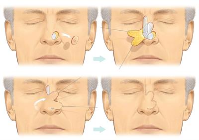
In the diagram above’s case, a skin cancer has been removed from the nose leaving a defect that cannot simply be stitched up. A local flap, called a bilobed flap is used to close the defect. The flap consists of skin along with the underling soft tissue and its blood supply.
A local flap like this relies on the fact that the skin has some natural elasticity and uses the lax skin in the bridge of the nose to close a defect near the tip of the nose where the skin is naturally tight.
Regional Flap

Uses a section of tissue that is attached by a specific blood vessel. When the flap is lifted, it needs only a very narrow attachment to the original site to receive its nourishing blood supply from the artery and vein.
This picture shows muscle from the calf being transferred, keeping its blood supply intact to cover an open fracture below the knee.
Free Flap / Microsurgery
Free flap reconstruction also involves the transfer of living tissue from one part of the body to another, along with the blood vessel that keeps it alive. A free flap is a further modification of flap transfer where the flap is entirely disconnected from its original blood supply and then reconnected using microsurgery in the recipient site. The following are two examples of free flaps.

In this image, the patient has an open fracture of his right lower leg. A muscle is taken from his inner left thigh and transferred to his right leg. The blood vessels that keep this muscle alive are dissected out of his left thigh along with the flap, divided, and then joined up micro-surgically to blood vessels in his right leg. This keeps the flap alive in its new position. To complete the reconstruction a split skin graft is taken from the left thigh and laid over the free muscle flap. This is called a free gracilis muscle flap.
This procedure involves hooking up the tiny blood vessels of the flap with those in the new site, and is carried out with use of a microscope, hence the name microsurgery.
The ability to disconnect and reattach tissue in this way means that the reach of flap is no longer confined by a patient’s anatomy.
More than any other technique, microsurgery has revolutionised plastic surgery as a specialty and is now the definitive treatment option for patients with cancer and major trauma.

In this example of a free flap a portion of skin and fat of the patient’s lower abdomen is transferred to the chest to reconstruct a breast. The blood vessels that keep this tissue alive are divided as they emerge from the groin area and rejoined micro-surgically to blood vessels in the chest in order to restore the blood supply to the flap. This is called a free TRAM flap breast reconstruction.
